Components & Functions
MRI machines vary in both size and shape. The older designs had a more
compact and small space and were very closed. This affected the patients mentally and
usually scared them even before they went in for the examination. However, the
engineers have tried to solve this problem by improving the machine to be more
open and inviting. They have expanded the sides and included much more space in
the scanner than the original models. The basic design of a Magnetic Resonance
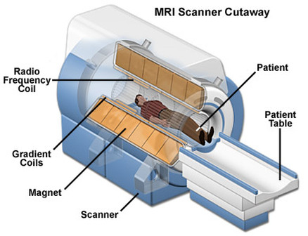
Imaging scanner is the same in almost all machines. The scanner consists of a 24
inch wide tube, inside which the examination takes place. It also contains a
magnet, a Radio Frequency (RF) coil, Gradient coils (3), patient table, and a
computer system.
compact and small space and were very closed. This affected the patients mentally and
usually scared them even before they went in for the examination. However, the
engineers have tried to solve this problem by improving the machine to be more
open and inviting. They have expanded the sides and included much more space in
the scanner than the original models. The basic design of a Magnetic Resonance
Imaging scanner is the same in almost all machines. The scanner consists of a 24
inch wide tube, inside which the examination takes place. It also contains a
magnet, a Radio Frequency (RF) coil, Gradient coils (3), patient table, and a
computer system.
Magnet
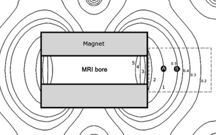
The magnet is the most important and biggest part of the MRI device. It is this
magnet that allows the MRI machine to produce high quality images. There is a
horizontal tube that runs through the magnet and is called a bore. The magnet
is extremely powerful and its strength is measured in either ‟teslaˮ or ‟gaussˮ
(1 tesla = 10 000 gauss). Most MRI magnets use a magnetic field of 0.5 to 2.0
tesla, when the Earth’s magnetic field is only 0.5 gauss. The magnetic field is
produced by passing current through multiple coils that are inside the magnet,
resulting in a state of superconductivity, which produces a lot of energy by
reducing the resistance in the wires to zero.
Gradient Coils
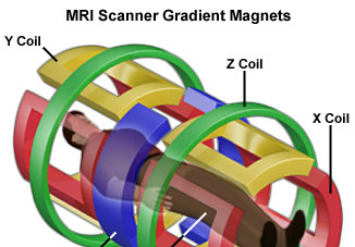
There are three different gradient coils
that are inside the MRI machine and are located within the main magnet. Each one
of these produce three different magnetic fields that are each less strong than
the main field. The gradient coils create a variable field (x, y, z) that can be
increased or decreased to allow specific and different parts of the body to be
scanned by altering and adjusting the main magnetic field.
Radio Frequency (RF) coils
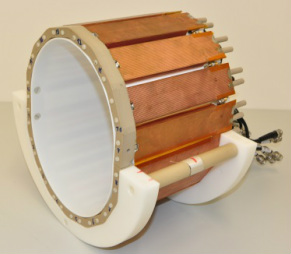
The basic function of the RF coils is
to transmit radio frequency waves into the patient’s body. There are different
coils located inside the MRI scanner to transmit waves into different body
parts. If a certain area of the body is specified, then all the RF coils usually
become focussed on the body part being imaged to allow for a better scan.
Patient Table
This component simply slides the patient into the MRI machine. The position at which
the patient lies down on the table is determined by the part of the body that is
being scanned. Once the part of the body under examination is in the exact
centre of the magnetic field, which is referred to as the isocentre, the
scanning process is started.
Antenna/Computer System
The antenna is a very sensitive device that easily detects the RF signals emitted by
a patient’s body while undergoing examination and feeds this information into
the computer system. The computer system is a powerful system, whose major
function is to receive, record, and analyze the images of the patient’s body
that have been scanned. It interprets the data sent in by the antenna and then,
helps to produce an understandable image of the body part being examined.

The Powerful MRI Computer System
