Brain MRI
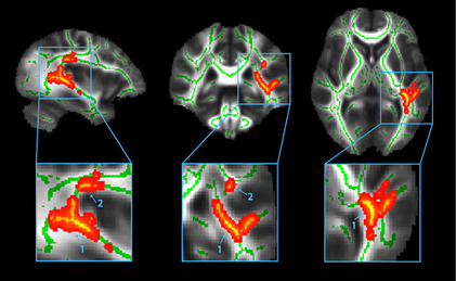
This diffusion tensor imaging scan depicts an united image of the fiber tracts which is also known as nerve fibers taken from 10 athletes with concussion and 10 athletes without. The three-dimensional scan clearly depicts the average image of every single white matter fiber tracts of the 20 samples. The green lines show the center of the fiber tracts. The areas of the brain that has been colored in red with yellow layering on top represents locations in which the concussed athletes show differences in their white matter fiber tracts. The bottom images zoom in on the two main areas where the differences were first observed.
Hand MRI
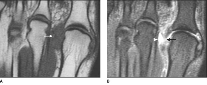
These two images show the MRI of two different patients left foot. Both of these patients have suffered from an acute ligament lesion, which is a paediatric disorder that includes ligament discontinuity, disinsertion (rapture), and thickening. The lesion has occurred in their
metacarpophalangeal joint that is located in the hand around the knuckles. As can be observed from image A, which is an axial MRI image of the first patient showing some fluid accumulation that has occurred in the soft tissues of the hand. This is indicated by the dark shade of gray to which the white arrow on the left is pointing to. In this area, obliteration (disintegration) of adjacent fats has also taken place, which
has led to the ligament lesion in the foot.
The second image, image B, which is a different kind of axial MRI scan performed on the second ligament lesion patient, shows the thickening of the ligament in between the
person’s knuckles. This is indicated by the black arrow to the right and this thickening of the ligament has caused a ligament discontinuity in the hand, which has resulted in a gap between the two joints in the kunckels because it’s our ligaments that hold our bones together with other bones and without them all the different joints and connections that we have between our bones would not exist. Furthermore, the white arrow head to the left is showing periligamental edema, which is a type of ligament haemorrhage, and the production
of some intra-articular fluid.
Knee MRI
This is an MRI scan of the mechanism of the knee. Figure 'A' shows the side view of the knee, while figure 'B' shows the knee from a bird's eye view. The arrow A pointing towards a tendon. Arrow B is also a tendon called the Patellar. The patellar is commonly known as the kneecap. Area C is where the Patella is and area D is the femur. Arrows E and F are cartilage muscles that help support the knee. Arrow H shows an insertion of the tendon. Knees have muscle tissues which allows them to help us move since the main function of muscle tissues is for movement.
