Scanning Process & Steps
There are multiple steps to completing a MRI scan. This process has to be carefully conducted and all factors must be controlled in order to achieve a usable, readable, high quality image. The MRI scanning process is concerned mostly with positively charged atoms, also known as protons. The human body consists of billions of atoms, however, since our body has a lot of water, which has a lot of positive hydrogen atoms in it, during the examination process these are the atoms that respond and are affected the most.
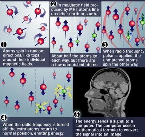
To begin with, the hydrogen atoms in the patient’s body are generally spinning in all directions on their imaginary axis, around their individual magnetic fields. This process is called precessing.
Next, when the patient is placed on the patient table and is inserted into the machine, the magnetic field of the scanner is turned on. The field that is produced causes the randomly spinning hydrogen particles to all line up in a strong magnetic moment. This begins the "magnetic" portion of the device. Since, the magnetic field runs right down the middle of the scanner, the atoms line up either facing north, which means they are aligned with the magnetic field, or facing south, which means they are anti-aligned with the magnetic field. During this step, about half of the atoms are expected to face north and half of atoms are expected to face south. However, when there is a damaged tissue, the atoms in that tissue remain unmatched and so, they don’t line up evenly as they are supposed to.
Steps of MRI Scanning
After this, a radio frequency pulse is applied by the RF coils to a specific area of the body that is being examined. This pulse is usually specific only to the hydrogen atoms, since those are the atoms that are abundant in our body. As the pulse is applied, all the atoms absorb it, however as the energy is taken in by the unmatched atoms, they end up turning around and facing the opposite direction to which they were previously spinning. This process is called "resonance" and depending on the force of the RF pulse, it is determined in which direction and at which rate the unmatched atoms spin. This frequency of resonance is called the Larmour Frequency.
While the RF pulse causes resonance to take place, at the same time the gradient coils are put into work. The three gradient coils- x, y, z- are turned on and off rapidly in a specific manner and their magnetic fields are also constantly increased and decreased. This allows for the observer to alter and focus the main magnetic field to a specific area, called the slice, and record as much detail as required in order to identify the problem.
When the RF pulse is finally turned off, the unmatched hydrogen atoms gradually return to their original position in relation to the main magnetic field. However, as they do so, these atoms, often classified as abnormal particles, emit a certain kind of energy.
This energy is then detected by the highly sensitive antenna, which feeds the data into the computer system in the form of waves or signals. Finally, the computer system interprets this data and converts the signal into a visible and understandable image that can be read and studied by the doctors and scientists.
